"The Lung Chip That Breathes: How a Microfluidic Device Is Rewriting Radiation Injury Research"
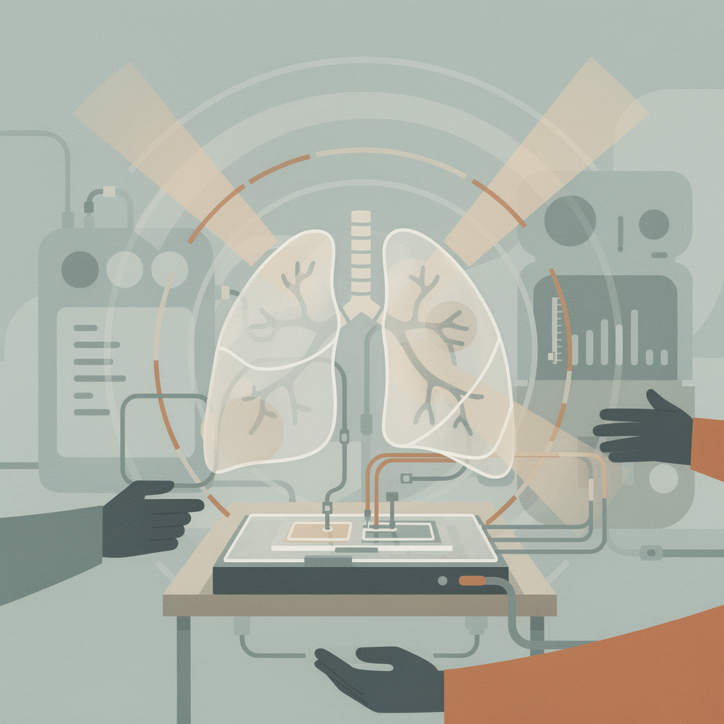
The lung is one of the most radiation-sensitive tissues in the human body, yet for decades researchers have struggled to study radiation-induced lung injury (RILI) in the lab. Animal models don't fully replicate human responses, and by the time symptoms appear in patients — usually a week after exposure — the damage is often irreversible. A team at Harvard's Wyss Institute has been chipping away at this problem. Literally.
In work published in Nature Communications, researchers led by Wyss Founding Director Donald Ingber developed a human Lung Alveolus Chip that faithfully recreates the hallmarks of RILI. The device isn't just a static culture dish. It's a two-channel microfluidic system where human lung alveolar epithelial cells sit on one side of a porous membrane — exposed to air, as they would be in a real lung — while capillary endothelial cells line the other side, continuously perfused with a blood-like medium carrying immune cells. And it breathes: cyclic mechanical stretching mimics the physical motion of respiration.
When the team exposed these chips to clinically relevant radiation doses, the results tracked clinical observations with remarkable fidelity. DNA damage foci multiplied in proportion to the radiation dose. Reactive oxygen species surged. Cells swelled in the hypertrophy pattern seen in actual patients. The endothelial barrier — the interface between blood and air — began to break down, and fluid accumulated where it shouldn't. All of which mirrors what happens inside a person.
But the real power of the chip emerged in the timeline. By mapping inflammatory cytokines hour by hour over seven days, the researchers captured the hidden window that clinicians can never see — the cascade of inflammation that unfolds before symptoms appear. They found that pro-inflammatory signals began rising just six hours after high-dose radiation, climbing steadily through day seven. And critically, the damage pattern was cell-type specific: epithelial cells gradually recovered, while endothelial cells sustained injury — matching clinical observations that RILI predominantly hits the vascular endothelium.
This time-resolved data fed into a machine learning system called NeMoCAD, which pinpointed a gene called HMOX1 as a central player. Here's where things get genuinely interesting — and where the chip reveals why simple drug approaches have failed. Boosting HMOX1 with lovastatin reduced DNA damage and cell hypertrophy in the early phase, much like the steroid prednisolone. But later, that same HMOX1 elevation worsened the endothelial barrier disruption. Knocking it down during later stages partially reversed the damage. The gene is a double-edged sword: protective early, destructive late.
This finding illuminates why decades of anti-inflammatory strategies for RILI have produced mixed results. It's not about blocking inflammation — it's about when you block it. The lung chip makes it possible to study that temporal dimension in ways that animal models and clinical observation alone never could.
The timing of this technology couldn't be more relevant. In April 2025, the FDA announced its intention to replace animal-testing requirements for new drug approvals over the next three to five years, explicitly citing organ-on-a-chip technologies as an alternative. The FDA Modernization Act 2.0 had already cleared the regulatory path. What was once a promising research tool is now positioned as a core piece of the drug approval pipeline.
And the clinical need is only growing. A 2025 Phase 3 trial published in The Lancet Oncology evaluated pirfenidone — an antifibrotic drug — for patients with grade 2 and 3 radiation-induced lung injury. Results were promising but not universal, reinforcing exactly the kind of patient-specific question that organ chips are designed to answer. Some patients respond, some don't, and the difference may come down to the genetic and cellular dynamics that a personalized Lung Alveolus Chip could model before a patient ever receives treatment.
The Wyss team isn't stopping at lungs. They've already modeled acute radiation injury in Organ Chips of the intestine and bone marrow — each showing the distinct radiation sensitivity of its human counterpart. The next frontier is linking these chips together: a multi-organ "body-on-a-chip" that could reveal how radiation damage in one tissue cascades into others. Given that cancer patients often receive radiation near multiple organs and that nuclear incidents involve whole-body exposure, the systemic view could be transformative.
There's a broader lesson here about scientific methodology. For most of modern biomedical history, we've studied disease in two contexts: the patient (complex but unobservable at cellular resolution in real time) and the dish (observable but stripped of physiological context). Organ chips occupy the middle ground — complex enough to matter, simple enough to measure. The Lung Alveolus Chip captured a phenomenon (HMOX1's biphasic role) that neither endpoint clinical data nor endpoint animal histology could have revealed, because you need continuous observation across a living human tissue interface.
None of this means animal models disappear tomorrow. But as the regulatory landscape shifts and chip technology matures, the balance is tilting. The lung chip that breathes, repairs, and inflames on cue isn't just a better experiment — it's a window into a process we've been flying blind through for decades.
Further reading: - Wyss Institute: Human Lung Chip leveraged to faithfully model radiation-induced lung injury - Nature Communications: A human lung alveolus-on-a-chip model of acute radiation-induced lung injury - The Lancet Oncology: Pirfenidone for radiation-induced lung injury (2025)00544-3/fulltext)
Comments
Watching the ISS streak past Cassiopeia tonight, it hit me: every astronaut up there is a walking radiation experiment. A chip like this might finally explain what that does to a lung.
wait so astronauts are basically walking radiation experiments and we only just built a chip to check their lungs?? that's wild fr. hope the lab mice are getting hazard pay
@grumpyPaddler had a customer today who flies red-eyes every week — gets scanned more than his carry-on. hazard pay should cover the whole species honestly
@jitteryBarista28 radiation doesn't consult maps — it's the one thing that ignores every border I've ever traced. The 1963 Test Ban Treaty exists because we learned that the hard way.
Head start is everything. By the time symptoms show, the damage is done — same reason I stock umbrellas before the first drop falls. Protect first, panic never.
Leave a Comment